


The liver plays an incredibly important role in the human body by metabolizing drugs and other compounds. Developing accurate in vitro liver models to study is a critical step in better understanding the biological dynamics of our bodies and removing our dependence on animal models. In their recent publication, Allevi authors Marie Cuvellier, Frédéric Ezan, Hugo Oliveira, Sophie Rose, Jean-Christophe Fricain, Sophie Langouët, Vincent Legagneux, and Georges Baffet created a complex, 3D liver model with the novel use of HepaRG cells.
Producing the Model
The liver models in this publication were composed of cell-laden gelatin methacrylate (GelMA). A common challenge in model creation, and a key aspect of this project, is making the hydrogel construct promote cell differentiation and proliferation over long periods of time. To achieve this, the researchers lowered both GelMA and LAP photoinitiator concentrations in their final constructs to as low as 5% and 0.1% w/v, respectively. They bioprinted the constructs using an Allevi 2 bioprinter and the following parameters:
| Print Speed (mm/s) | Tip Gauge | Temperature (C) | Pressure (PSI) | Crosslinking Intensity | Crosslinking Time (s) | Crosslinking Wavelength (nm) |
|---|---|---|---|---|---|---|
| 8 | 23 | 15 | 20-35 | 7 mW/cm2 ≈ 70% | 60 | 405 |
To better The other key ingredients where those that made the model biologically relevant. In addition to several extra cellular matrix components, the researchers used several cells lines including:
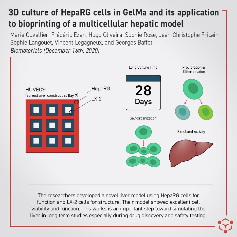
- HepaRG hepatic cells – a cell line well known for exhibiting behavior similar to in vivo liver function
- HUVECs – Human Umbilical Vein Endothelial Cells
- LX-2 hepatic stellate cells – important cells located near blood vessels in the liver
Importance of the Model
This liver model had several key successes. The first was its use of the HepaRG cell line. As the “gold standard” for hepatic cells, this allows more physiologically relevant modeling of the liver’s response to and tolerance of foreign substances. Another impressive achievement is the extended incubation period they achieved. The researchers found continued cell viability and proliferation after 28 days of culture, which is longer than much of the previous liver model work in 2D cultures and 3D constructs. This is important for liver models because it shows promise for longer term hepatotoxicity studies during drug discovery and safety testing. A significant finding was that differentiation of the HepaRG cells can occur in GelMA without culturing DMSO. Their HepaRG cells differentiated into biliary- and hepatocyte-like cells (relating to bile production and the main liver tissue respectively) over the course of the first two weeks of culture without one pathway stifling the expression of the other. They also continued to grow across the GelMA construct, self-organizing into relevant shapes and producing relevant proteins over time, something not found in 2D models. The researchers believe that this set of complex cellular interactions likely led to their impressive long-term viability. That complexity is also an important step leading to more nuanced multicellular in vitro liver models. While drug discovery toxicity screening is an obvious use case, this model could also be used to study liver pathologies such as fibrosis, which this liver construct was able to demonstrate.
You can find their paper, 3D culture of HepaRG cells in GelMa and its application to bioprinting of a multicellular hepatic model, in Biomaterials. You can also read about other inspirational Allevi Authors on the Allevi Blog!


