Allevi Blog
Allevi Author: Lattices vs Sheets for Cardiac Tissue Bioprinting
- Updated on April 3, 2020
There are so many variables that go into creating viable 3d bioprinted tissues; bioink selection, print geometry, cure times, rigidity, flexibility, degradation time and cell viability to name a few. Not to mention, each of these parameters needs to be analyzed and perfected for every cell line in the body. As a community, we are still figuring out the perfect protocol for each organ system.
In a new paper out this week titled “A Comparative Study of a 3D Bioprinted Gelatin-Based Lattice and Rectangular-Sheet Structures”, our newest Allevi Authors tackled one of these lingering questions, “What is the best print structure for cardiac tissue when comparing lattices vs sheets?”
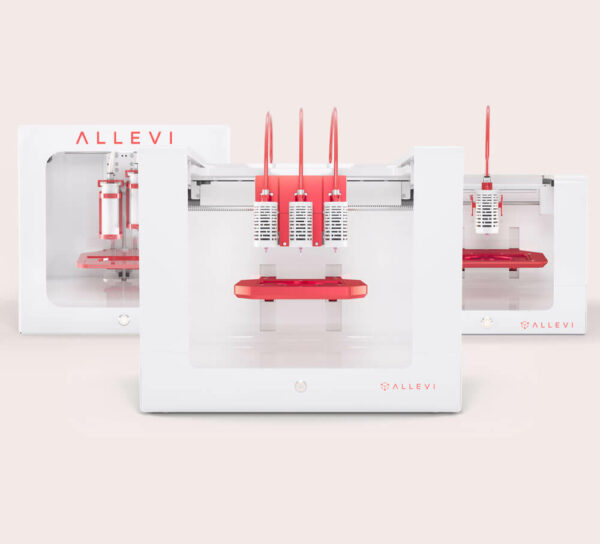
Researchers at University of Texas El Paso and University of Texas at Austin used their Allevi 2 bioprinter and furfuryl gelatin to study and compare 3d bioprinted lattices vs sheets. Through their comparison, they discovered that the lattice structure was more porous with enhanced rheological properties and exhibited a lower degradation rate compared to the rectangular-sheet.

Further, the lattices allowed cells to proliferate to a greater extent compared to the rectangular-sheets. All of these results collectively affirmed that lattices pose as a superior scaffold design for tissue engineering applications.
Read the full paper here to learn more about the rigorous testing and analysis the team conducted during their study.

