Allevi Blog
Allevi Author: Tohoku University Imaging 3D Hydrogels
- Updated on March 31, 2020

One of the most rewarding “AHA” moments of bioprinting is seeing your cells proliferate within a 3D tissue. As 3D bioprinting becomes more widely adopted within the fields of tissue engineering and personalized medicine, it is important that researchers are able to monitor cell activity within a 3D structure. Our most recent Allevi Authors from Tohoku University developed a method for imaging 3D hydrogels by electrochemically monitoring a tissue.
Their paper, titled “Electrochemical Imaging of Cell Activity in Hydrogels Embedded in Grid-Shaped Polycaprolactone Scaffolds Using a Large-scale Integration (LSI)-based Amperometric Device”, was published in the Analytical Sciences Journal.
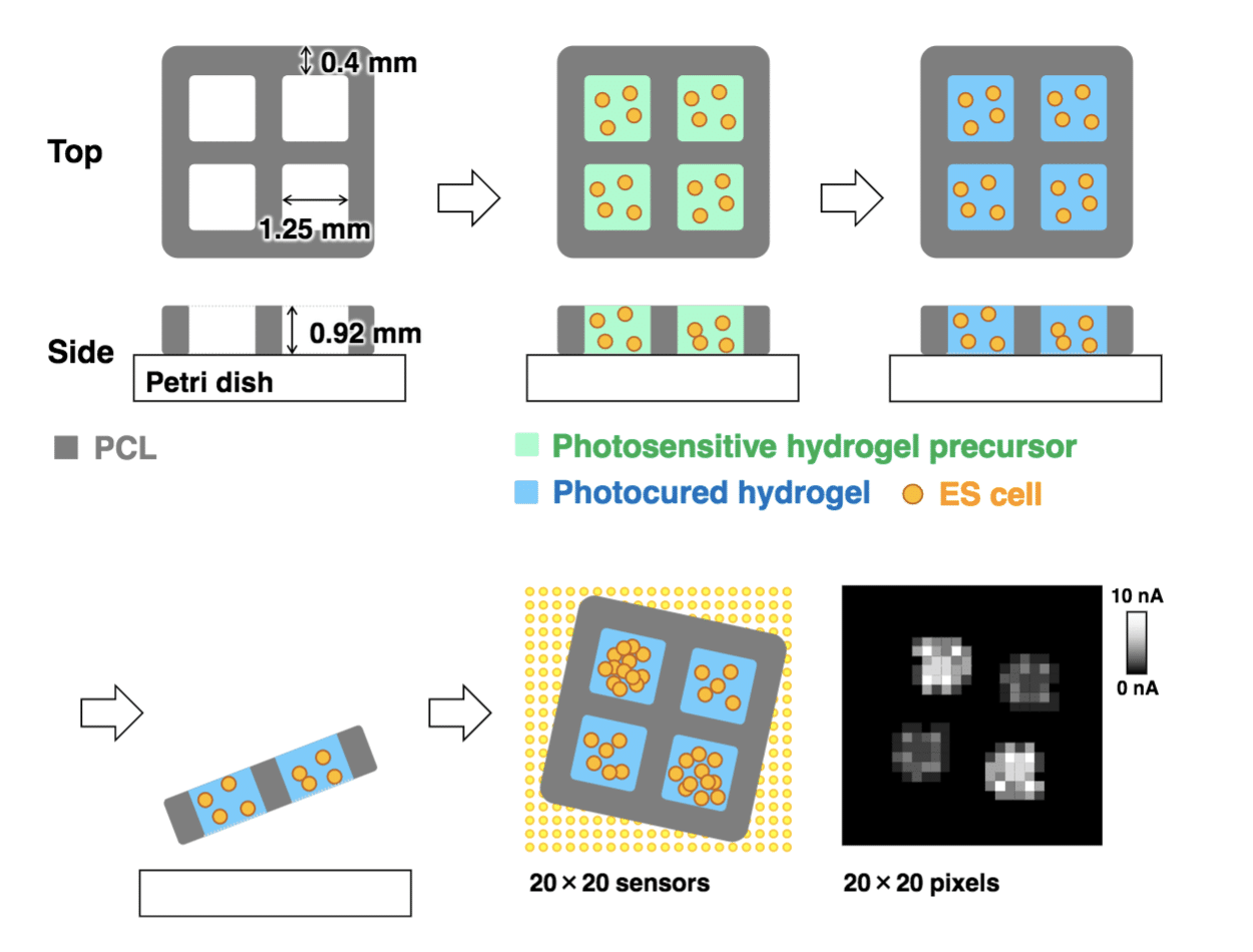
Tissue engineering requires analytical methods to monitor cell activity in hydrogels. Here, we present a method for the electrochemical imaging of cell activity in hydrogels embedded in printed polycaprolactone (PCL) scaffolds. Because a structure made of only hydrogel is fragile, PCL frameworks are used as a support material. A grid-shaped PCL was fabricated using an excluder printer. Photocured hydrogels containing cells were set at each grid hole, and cell activity was monitored using a large-scale integration-based amperometric device. The electrochemical device contains 400 microelectrodes for biomolecule detection, such as dissolved oxygen and enzymatic products. As proof of the concept, alkaline phosphatase and respiration activities of embryonic stem cells in the hydrogels were electrochemically monitored. The results indicate that the electrochemical imaging is useful for evaluating cells in printed scaffolds.
Tohoku University
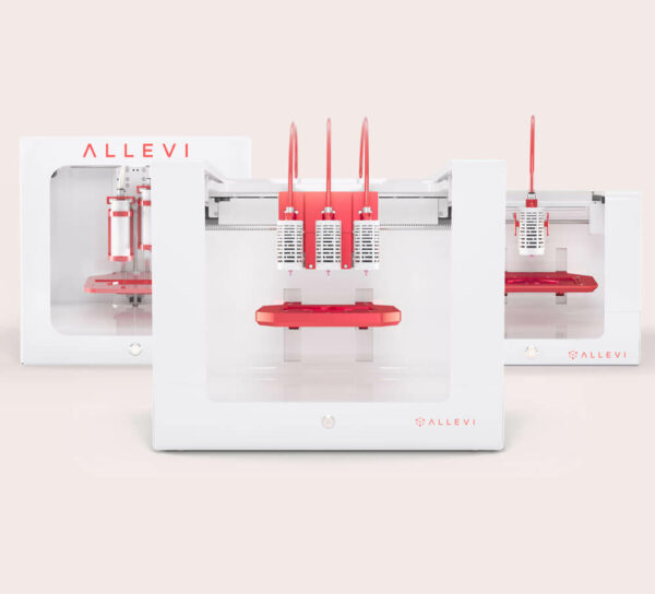
The researchers from Tohoku University use their Allevi 2 bioprinter to print PCL scaffolds as a support material for photocured hydrogels. They then used an amperometric device to electrochemically monitor the living cells. Through their study, they were able to determine that electrochemical imaging is a great way to monitor cell differentiation and will be useful for evaluating the viability of thicker bioprinted tissues.
Congratulations to the Tohoku University researchers on their findings for imaging 3D hydrogels!

